Canthaxanthin Toxicity and Membrane Damage: Why Tanning Pills Failed
Canthaxanthin, known chemically as beta, beta-carotene 4, 4' dione, has a history of widespread use as an ingestible drug, food colorant, and cosmetic colorant. In past decades, the compound attracted attention among consumers seeking a bronzed appearance through oral tanning tablets. However, systemic intake exposed unexpected hazards, leading medical professionals to abandon endorsing canthaxanthin for cosmetic coloring. The emergence of ocular complications prompted researchers to investigate the underlying cellular mechanisms responsible for tissue injury. A key benchmark in this scientific journey is a 2009 review in Cellular & molecular biology letters, which synthesized laboratory evidence on how canthaxanthin disrupts the delicate architecture of lipid membranes.
Background: From Cosmetic Colorant to Systemic Concern
The journey of canthaxanthin illustrates the significant divide between consuming a color additive in minor amounts and utilizing systemic agents to alter complexion. When ingested orally, the compound distributes through human tissues rather than remaining on the skin surface. Unlike topical coloring agents discussed in guides on safer alternatives to melanotan, ingested pigments pass through the bloodstream and interact directly with cellular structures throughout the body. Over time, reports revealed that canthaxanthin produces undesirable effects on human health, with the most pronounced damage localized in the eyes.
The Clinical Concern: Macular Crystals and Vascular Injury
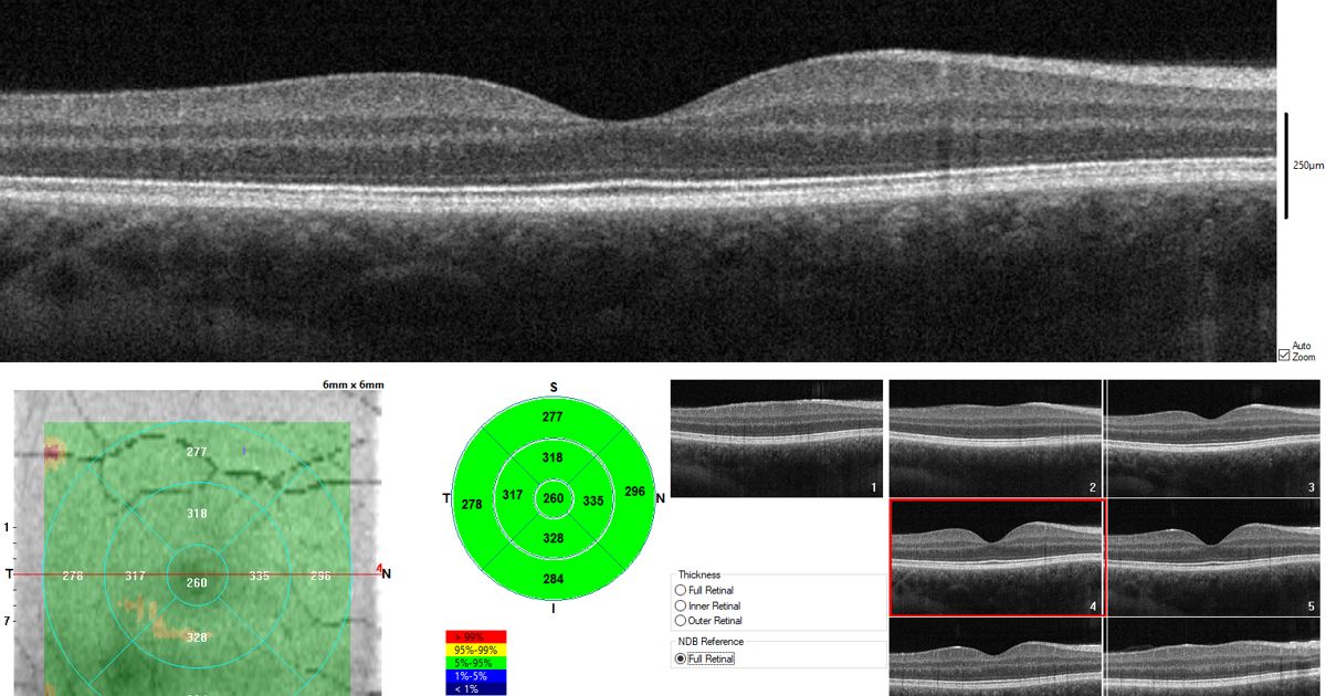
The primary medical issue tied to systemic canthaxanthin consumption is an eye condition termed canthaxanthin retinopathy. This adverse condition is defined by the distinct formation of crystals within the macula lutea membranes of the human retina. The macula is responsible for high-acuity central vision, making any foreign accumulation a critical threat to sight. Clinical examinations established that canthaxanthin retinopathy does not merely present benign physical deposits; the eye dysfunction is strongly connected with objective damage to the delicate blood vessels surrounding the exact location of crystal deposition. Recognizing this vascular harm caused medical professionals to move away from systemic canthaxanthin administration.
Hypotheses on Toxicity: Aggregation and Lipid Interactions
To explain why crystals form and injure nearby vessels, scientific investigators proposed that the molecular properties of the carotenoid itself are responsible. The working hypothesis evaluated in the literature suggests that direct interactions between canthaxanthin and cellular lipid membranes, alongside the physical aggregation of the pigment, serve as primary drivers of vascular toxicity. When individual pigment molecules cluster together, they alter the surrounding cellular environment, predisposing the retinal microvasculature to dysfunction and structural breakdown.
Inside the 2009 Review: Examining Experimental Membrane Models
To assess these biological interactions without risking patient safety, scientists examined laboratory model systems. As detailed in the published paper Interactions between canthaxanthin and lipid membranes--possible mechanisms of canthaxanthin toxicity, researchers reviewed experimental data gathered from multiple simplified membrane structures:
- Monolayers composed of pure canthaxanthin
- Mixed monolayers containing both canthaxanthin and lipids
- Oriented lipid bilayers
- Artificial liposomes modeled to reflect biological barriers
Across every model system tested, the experimental results showed a very strong effect of canthaxanthin on the fundamental physical properties of lipid membranes. The data indicated that incorporating this pigment modifies the flexibility, arrangement, and stability of lipid layers, providing a plausible biophysical explanation for how canthaxanthin exerts toxic actions that lead to progressive canthaxanthin retinopathy.
Study Design and Scientific Limits
While the findings offer meaningful insight into cellular mechanics, it is important to interpret this work within its proper scientific context. This evidence comes from a single review paper that focused on in vitro model systems, such as liposomes and lipid monolayers, rather than dynamic living human organs. These controlled laboratory platforms demonstrate how the molecule alters physical membrane properties in isolation, but they do not capture the complete biological complexity of living retinal tissue, metabolic variations, or clearance mechanisms across diverse individuals. For this reason, the findings illustrate a plausible toxic mechanism rather than an exhaustive proof of every systemic effect, reinforcing why consumers must always discuss pigment-altering chemicals and melanotan risks and side effects with a certified dermatologist or medical doctor.
Medical Perspective on Tanning Substances
The clinical abandonment of canthaxanthin demonstrates why oral and systemic shortcuts to skin pigmentation carry serious risks. Whether reviewing past tanning tablets or modern unregulated substances like melanotan sold through online shops, altering cellular biology without regulated medical oversight poses unpredictable dangers to organ systems. Anyone experiencing vision changes or considering systemic products intended to change skin color should consult an eye specialist or dermatologist to protect their long-term health.
Sources
Published October 31, 2026. Sources are linked in the text. See our Medical Disclaimer.