Comparing Surgical Excision and Removal for Localized Skin Cancer
When an abnormal skin growth or suspicious lesion appears, medical evaluation focuses on determining whether malignant cells are present and physically removing the diseased tissue. For localized nonmelanoma growths such as basal cell carcinoma and squamous cell carcinoma, surgical removal and precise biopsy procedures represent the primary standard of clinical care. Deciding on the appropriate removal method depends on how deeply abnormal cells extend into the cutaneous layers, the tumor's physical size, and its location on the body. Understanding how dermatologists and surgeons evaluate, sample, and excise localized lesions provides critical clarity for anyone reviewing standard dermatological procedures.
How Localized Cancers Form Across Skin Layers
The skin serves as the body's largest organ, providing protection against heat, sunlight, injury, and infection while storing water, fat, and vitamin D. It consists of two main layers: the outer epidermis and the inner dermis. Skin cancer originates within the epidermis, which is made up of three kinds of cells: thin, flat squamous cells forming the top surface, round basal cells situated beneath them, and melanocytes residing in the lower section.
Melanocytes produce melanin, the pigment responsible for skin color, which increases when exposed to natural or artificial ultraviolet light. Uncontrolled malignant transformation among these cell types leads to specific conditions, as outlined in our review of types of skin cancer. Basal cell carcinoma and squamous cell carcinoma represent nonmelanoma skin cancers. In addition, precancerous changes such as actinic keratosis can emerge as rough, scaly patches on sun-exposed skin, which can sometimes advance into squamous cell carcinoma if left untreated.
Comparing Excisional Removal and Biopsy Modalities
According to the National Cancer Institute's skin cancer treatment guide, confirming whether an abnormal growth is malignant requires tissue removal for microscopic examination by a pathologist. Physicians utilize four main types of skin biopsies depending on the size and location of the lesion:
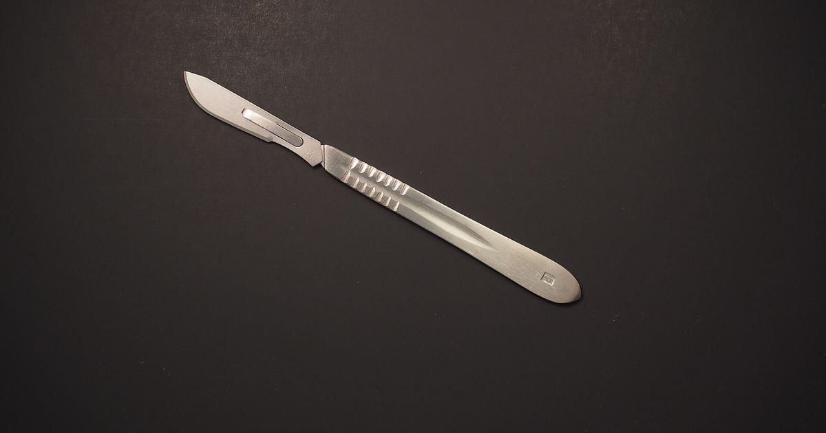
- Excisional biopsy: A surgeon or dermatologist uses a scalpel to remove the entire growth from the skin. This approach eliminates the visible tumor while providing complete tissue margins for laboratory study.
- Incisional biopsy: A scalpel is used to remove only a portion of the abnormal growth when complete immediate removal is not practical.
- Punch biopsy: The doctor uses a circular instrument called a punch or trephine, rotating it clockwise and counterclockwise to cut about 4 millimeters down into the layer of fatty tissue beneath the skin to extract a round sample.
- Shave biopsy: A sterile razor blade is used to shave off the abnormal-looking growth from the upper layers of the skin.
Pathologic Staging and Tissue Margin Evaluation
When surgery is performed to remove a primary tumor along with abnormal lymph nodes, the excised tissue is studied under a microscope. As detailed by the National Cancer Institute, this process is known as pathologic staging. Pathologic findings offer definitive structural information that may differ from clinical staging, which relies on physical examinations and imaging conducted prior to surgical intervention.
Pathologic analysis establishes whether cancer cells remain strictly localized or have expanded. For nonmelanoma skin cancer on the head or neck, formal stages guide clinical action. In Stage 0, also referred to as carcinoma in situ, abnormal cells exist exclusively within the squamous or basal cell layer of the epidermis and have not invaded nearby normal tissue. Once the disease progresses to Stage I, a distinct tumor has formed that measures 2 centimeters or smaller. Complete surgical excision aims to clear these localized tumors before malignant cells penetrate surrounding anatomical structures.
Clinical Factors Influencing Surgical Decisions
Choosing an appropriate procedural strategy requires balancing complete oncologic clearance with functional preservation. Doctors evaluate several specific clinical factors before determining how to manage localized disease:
- Cancer classification: Basal cell carcinoma rarely spreads to distant regions of the body, meaning extensive staging tests are usually unnecessary. In contrast, squamous cell carcinoma carries a higher risk of local invasion and metastasis.
- Anatomical site and size: Lesions appearing on sun-exposed areas such as the face, neck, hands, nose, ears, or lower lip require meticulous surgical borders.
- Patient health characteristics: A patient's overall general health, tobacco use, and whether the immune system is weakened significantly alter prognosis and guide procedural planning.
How Localized Disease Spreads if Left Unchecked
When localized skin cancers are not diagnosed or removed in a timely manner, malignant cells can advance through three distinct mechanisms: by direct invasion into nearby tissue, by entering the lymph system and traveling through lymph vessels, or by entering the bloodstream and moving through blood vessels. When cancer forms a metastatic tumor in another organ, the newly established tumor consists of the original skin cancer cells. To assess whether squamous cell carcinoma has traveled, physicians may perform computed tomography scans, chest x-rays, positron emission tomography scans using injected radioactive glucose, or ultrasound examinations of regional lymph nodes.
Sources
Published October 30, 2026. Sources are linked in the text. See our Medical Disclaimer.