Cutaneous Porphyrias and Phototoxicity: Key Clinical Diagnostics
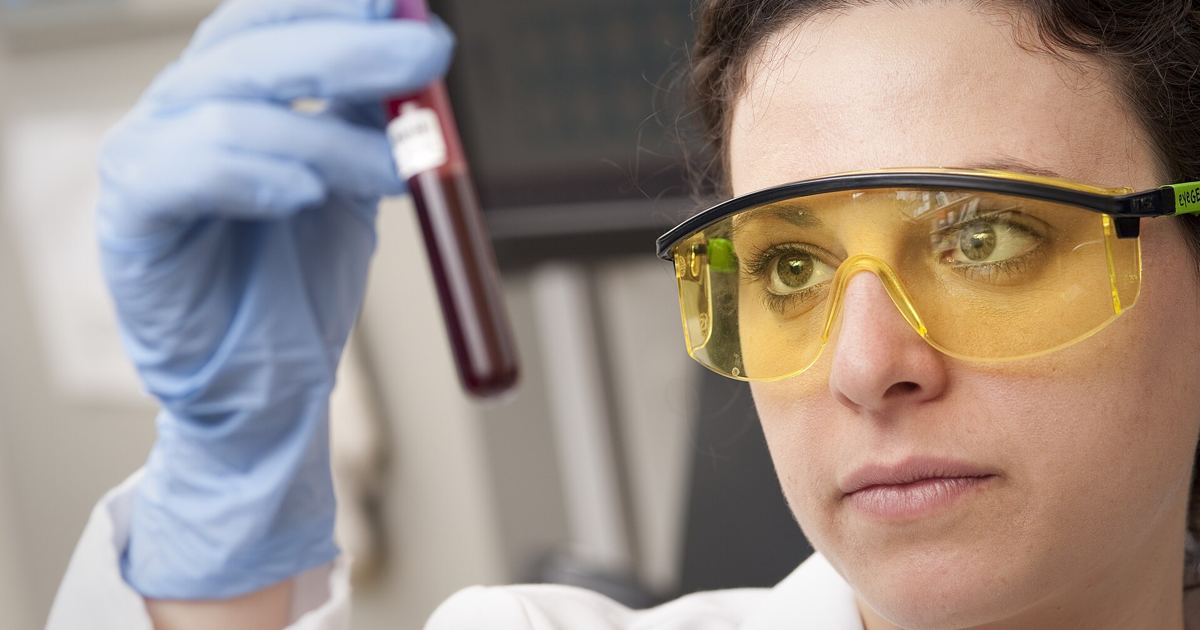
Severe, unexpected phototoxic reactions can occur when underlying metabolic pathways are disrupted. Among the most dramatic causes of acute light intolerance are porphyrias—complex metabolic disorders resulting from enzyme deficiencies within the heme biosynthetic pathway. According to a 2019 review titled Clinical Guide and Update on Porphyrias. published in Gastroenterology, identifying these rare conditions requires targeted biochemical testing to separate true porphyric disorders from secondary elevations and to guide appropriate medical care.
Study Design and Methodological Limits
The 2019 paper in Gastroenterology is a narrative clinical review rather than an original observational cohort or randomized trial. As a single clinical guide, it consolidates diagnostic criteria, enzymatic pathways, and therapeutic interventions across both acute and non-acute forms of the disorder. A key limitation of this review format is that it relies on existing literature and cannot provide new statistical prevalence data across broader populations. Readers should view the findings as a synthesized clinical guide to heme pathway dysfunction rather than absolute outcomes across every clinical demographic.
The 2 Overarching Categories: Acute vs. Non-Acute
The heme biosynthetic pathway can fail at various stages, creating distinct clinical profiles. Clinicians divide porphyrias broadly into two primary operational categories:
- Acute Hepatic Porphyrias: This group includes acute intermittent porphyria, variegate porphyria, hereditary coproporphyria, and aminolevulinic acid dehydratase deficient porphyria. These disorders manifest as sudden attacks driven by an overproduction of early porphyrin precursors. Symptoms can involve severe abdominal, neurologic, psychiatric, or cardiovascular complications. Among these, variegate porphyria and hereditary coproporphyria can also cause acute skin photosensitivity.
- Non-Acute Porphyrias: This category encompasses porphyria cutanea tarda, erythropoietic protoporphyria (EPP), X-linked protoporphyria, and the rare congenital erythropoietic porphyria. Instead of producing neurovisceral attacks, these disorders lead to an accumulation of porphyrins that trigger severe cutaneous photosensitivity and, in some cases, serious liver damage.
Key Diagnostic Markers: Precursors vs. Porphyrins
Diagnosing these conditions requires specific biochemical testing rather than standard blood counts. For acute hepatic porphyrias, initial assessment relies heavily on quantifying urinary porphyrin precursors:
- In aminolevulinic acid dehydratase deficient porphyria, clinicians specifically measure increased levels of urinary 5-aminolevulinic acid.
- In the remaining acute porphyrias, diagnostic testing evaluates both elevated urinary 5-aminolevulinic acid and increased porphobilinogen.
In non-acute cutaneous porphyrias, testing focuses on elevated porphyrins accumulating in the blood, urine, or tissues, which directly provoke phototoxic crises when skin meets sunlight.
3 Confounding Factors in Differential Diagnosis
A critical lesson from the Gastroenterology porphyrias guide is that elevated porphyrin levels do not automatically indicate an inherited enzymatic defect. Secondary elevations can confuse clinical interpretation. Physicians must rule out non-porphyria conditions that trigger secondary increases in porphyrins or precursors:
- Liver Disease: Underlying hepatic conditions can alter excretion pathways, causing secondary elevations in urinary or blood porphyrins.
- Iron Deficiency: Low iron stores can secondarily disrupt intermediate biochemical steps.
- Lead Intoxication: Exposure to lead directly inhibits metabolic enzymes, yielding increases in both porphyrins and porphyrin precursors.
Targeted Medical Interventions and Photoprotection
Management depends strictly on the exact enzymatic defect identified. For severe acute attacks, patients require intensive care, intravenous heme therapy, caloric support, and rigorous avoidance of porphyrinogenic drugs or triggers.
For porphyria cutanea tarda, treatment protocols include therapeutic iron depletion, hydroxychloroquine therapy, and the eradication of hepatitis C virus when present. For patients facing profound light sensitivity from erythropoietic protoporphyria or X-linked protoporphyria, the review highlights that an alpha-melanocyte-stimulating hormone analogue can significantly reduce sunlight sensitivity. Patients curious about this therapeutic class often research afamelanotide (Scenesse), the regulated, approved synthetic peptide developed for light intolerance, which stands in stark contrast to unregulated tanning products. Individuals taking prescription therapies should also review broader medical advice regarding drug-induced photosensitivity.
Anyone experiencing abnormal burning, redness, or cutaneous pain following sun exposure should immediately consult a board-certified dermatologist or primary care doctor for appropriate diagnostic laboratory testing.
Sources
- Clinical Guide and Update on Porphyrias. - Gastroenterology (PubMed PMID 31085196)
Published October 30, 2026. Sources are linked in the text. See our Medical Disclaimer.