DHA and Skin Cells: FAQ on the 2020 Stress Response Study
For individuals seeking cosmetic color without ultraviolet exposure, dihydroxyacetone (DHA) has long served as the central active ingredient in sunless tanning lotions and sprays. Many people explore these products as sunless tanning alternatives instead of unregulated injectable peptides. While chemical tanning avoids the solar radiation known to cause epidermal genotoxic stress, researchers continue to study how DHA interacts with cutaneous biology. A 2020 laboratory study published in Redox Biology investigated whether DHA triggers cellular stress responses in cultured human skin cells and laboratory tissue reconstructs. Below are answers to common questions regarding the study design, molecular observations, and research boundaries.
How does dihydroxyacetone create pigment on the skin?
Chemical tanning relies on producing pigments that mimic melanin, termed melanoidins. This process occurs through spontaneous amino-carbonyl reactions, also known as glycation. In this reaction, epidermal amino acid and protein components interact with reactive sugars, specifically the glycolytic ketose dihydroxyacetone (DHA). Because this coloration develops within the outer cutaneous layers without sunlight, it has widely been regarded as a safer substitute for solar exposure. However, the specific cellular biology impacted by this chemical tanning process had remained largely unexplored prior to detailed molecular investigations.
What did the 2020 study examine in cultured keratinocytes?
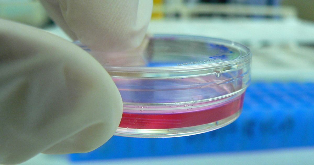
The investigators examined acute exposure to DHA using cultured human HaCaT keratinocytes. When exposed to low millimolar concentrations of DHA, the cells did not experience impaired viability. However, the researchers observed a pronounced cellular stress response. This response involved the rapid activation of phospho-protein signal transduction pathways, including p-p38, p-eIF2α, and p-Hsp27 at serine sites S15 and S78.
Alongside signal transduction changes, the keratinocytes demonstrated specific alterations in gene expression. Genes showing altered expression included HSPA6, HMOX1, CRYAB, and CCL3. To determine whether this reaction was unique to the active tanning agent, the scientists tested glycerol as a non-ketose, tanning-inactive control. Keratinocytes exposed to glycerol did not display these stress-signaling activations or gene expression changes.
Did the researchers detect advanced glycation end products?
Yes. Because DHA reacts through glycation, the study analyzed whether posttranslational protein modifications occurred. Using quantitative mass spectrometric detection, the researchers confirmed the formation of advanced glycation end products (AGEs). Specifically, they identified N-ε-(carboxyethyl)-l-lysine (CEL) and N7-carboxyethyl-l-arginine within the treated cells.
The study also evaluated how cells manage this chemical burden. When the scientists applied CRISPR-Cas9 gene editing to eliminate the carbonyl stress response gene GLO1, which encodes the enzyme glyoxalase 1, the modified skin cells showed hypersensitivity to cytotoxicity from DHA. This finding indicates that functional cellular defense pathways play a role in processing acute carbonyl stress caused by the compound.
What markers appeared in reconstructed human epidermis and animal models?
To move beyond single-cell layers, the research team applied a topical, use-relevant DHA regimen to human reconstructed epidermis. A gene expression array revealed a comparable stress response in these three-dimensional tissue models, including changes across multiple genes:
- Gene expression changes involving HSPA1A, HSPA6, and HSPD1.
- Signaling and transcriptional alterations affecting IL6, DDIT3, and EGR1.
- Immunohistochemical detection showing the presence of CEL, HO-1, and p-Hsp27-S78.
The researchers also evaluated SKH-1 hairless mouse skin following DHA treatment. Immunohistochemical analysis of the treated mouse tissue confirmed the epidermal occurrence of both CEL and p-Hsp27 epitopes, mirroring several cellular events observed in human tissue reconstructs.
How did the DHA response compare to ultraviolet radiation?
Solar ultraviolet exposure is well documented to cause direct epidermal genotoxic stress. In this laboratory study published in Redox Biology, the authors performed a comparison by exposing epidermis to a supra-erythemal dose of solar simulated UV, set at 2 MEDs. They used gene expression array analysis to track how tissue responded to each stimulus.
The comparative array revealed that certain stress response genes were equally sensitive to both DHA and solar UV, while other genes responded differentially to either chemical tanning or simulated sunlight. While both stimuli prompted measurable stress pathways in laboratory models, the mechanisms driving them differ fundamentally. Consumers researching tanning methods should also recognize that unapproved synthetic substances, detailed in educational overviews such as what is melanotan, carry distinct systemic concerns and lack regulatory clearance, making direct consultation with a doctor or dermatologist essential for personalized skin guidance.
What are the primary limitations of this research?
It is crucial to keep this research in perspective. This paper represents a single experimental study conducted in vitro on cell cultures, reconstructed tissue models, and hairless mice. It did not track health outcomes, systemic absorption, or chronic usage patterns in living human subjects. As the study authors emphasized, these findings represent prototype data documenting a specific cutaneous stress response that warrants further molecular exploration in living human skin rather than definitive clinical conclusions.
Sources
Published October 28, 2026. Sources are linked in the text. See our Medical Disclaimer.