Giant Congenital Pigmented Nevi: A Clinical and Monitoring Checklist
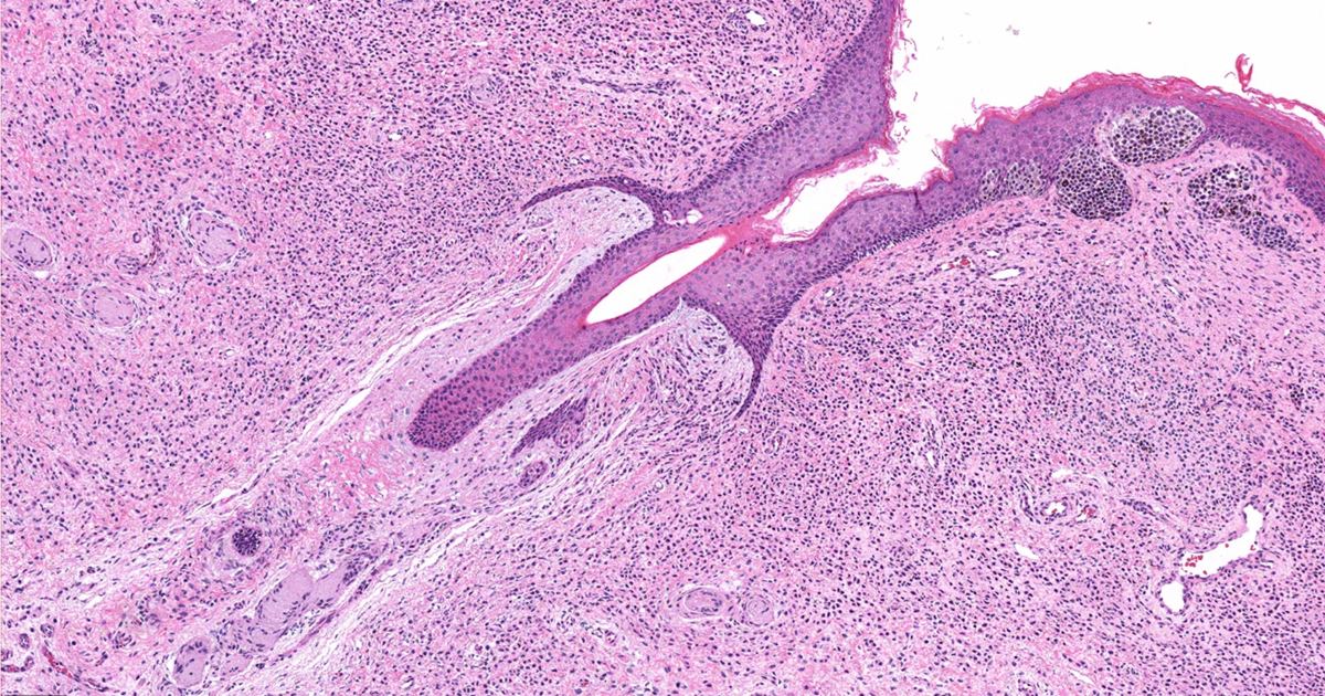
A congenital pigmented or melanocytic nevus is a dark-colored patch of skin that is present right at birth or appears during the first year of life. When these marks reach exceptional sizes, medical teams classify them as giant congenital nevi. Understanding how these marks form, what signs require close medical evaluation, and how healthcare providers monitor them over time can help families navigate ongoing skin care safely.
Understanding Giant Congenital Nevi and Causes
According to the MedlinePlus Medical Encyclopedia entry on giant congenital nevus, a congenital nevus contains an increased amount of melanocytes. Melanocytes are specialized skin cells that produce melanin, the pigment responsible for skin color. During fetal development in the womb, a localized increase in these melanocytes creates the pigmented patch. Medical experts believe this condition stems from a variant gene.
While smaller congenital nevi are common in children and do not cause problems most of the time, large or giant nevi are rare. In infants and children, a giant congenital nevus begins smaller, but it typically grows as the child grows. By the time it stops growing, a giant pigmented nevus is larger than 15 inches or 38.1 centimeters across.
Clinical Features Checklist: What to Look For
Healthcare providers look for specific physical characteristics when evaluating these birthmarks. You can review your child's skin alongside a doctor using this checklist of clinical features:
- Color: The lesion appears as a dark patch ranging from brown to bluish-black.
- Hair growth: The patch is often hairy (sometimes referred to as a giant hairy nevus).
- Borders: The edges can present with regular or uneven borders.
- Surface texture: The skin surface may feel smooth, irregular, or distinctly wart-like.
- Satellite marks: Smaller affected pigmented areas sometimes appear near the larger nevus.
- Body location: These marks commonly develop on the upper or lower parts of the back or on the abdomen. They can also occur on the arms, legs, mucus membranes, palms, or soles.
A giant nevus may also occur alongside other nevi across the body. When comparing dark marks or monitoring changes, exploring our guide on common moles vs. dysplastic nevi can provide additional dermatological context.
Medical Exams, Diagnostic Tests, and Spine Involvement
Because giant lesions involve deep layers of pigment cells, specialized medical evaluations are essential. Every birthmark should be looked at by a healthcare provider. Diagnostic and monitoring protocols may include:
- Annual measurements and medical photography: Your provider will measure the dark skin area every year and may take pictures to check if the spot is getting larger.
- Skin biopsy: A provider may perform a skin biopsy to confirm the clinical diagnosis or check for cancer cells.
- Spinal and brain imaging: An MRI of the brain or spine might be ordered if the nevus sits over the spine. A giant nevus over the spine may be linked to brain or spinal cord problems, and involvement of the membranes of the brain and spinal cord can occur when the nevus affects a very large area.
Evaluating Melanoma Risk and Complications
The primary medical concern with large congenital lesions is skin cancer. Skin cancer (melanoma) can develop in some people with large or giant nevi. Clinical data reviewed by MedlinePlus shows that the cancer risk is higher for nevi that are larger in size. You can learn more about how malignant changes arise in our review on melanotan, moles, and melanoma case reports.
Beyond skin cancer, having a giant nevus may lead to depression and other emotional problems if the birthmark affects appearance. Ongoing emotional support and open discussions with medical teams play an important role in long-term care.
Management and Treatment Options Checklist
Management of a giant pigmented lesion requires regular exams to check for skin cancer. Depending on the location, size, and health considerations, medical teams may discuss several management approaches:
- Surgical removal: Surgery can be done for cosmetic reasons or if your provider thinks the lesion may become skin cancer. Larger nevi may require removal in several stages, and skin grafting is done when needed. However, it is not currently known if removing the nevus reduces the overall skin cancer risk.
- Dermabrasion and lasers: Providers can use dermabrasion (rubbing the cells off) or lasers to improve physical appearance. However, these surface treatments may not remove the whole birthmark, which can make it harder to detect skin cancer later.
- Psychological consideration: Treatment can be helpful if the lesion causes emotional distress due to how it looks. Patients and families should talk directly with their provider about the pros and cons of surgery.
If you or your child has a large, pigmented area anywhere on the skin, consult a doctor or dermatologist promptly for an individualized monitoring schedule.
Sources
Published October 30, 2026. Sources are linked in the text. See our Medical Disclaimer.