Hair Follicle Pigmentation and mTORC1: Cellular Pathways Explained
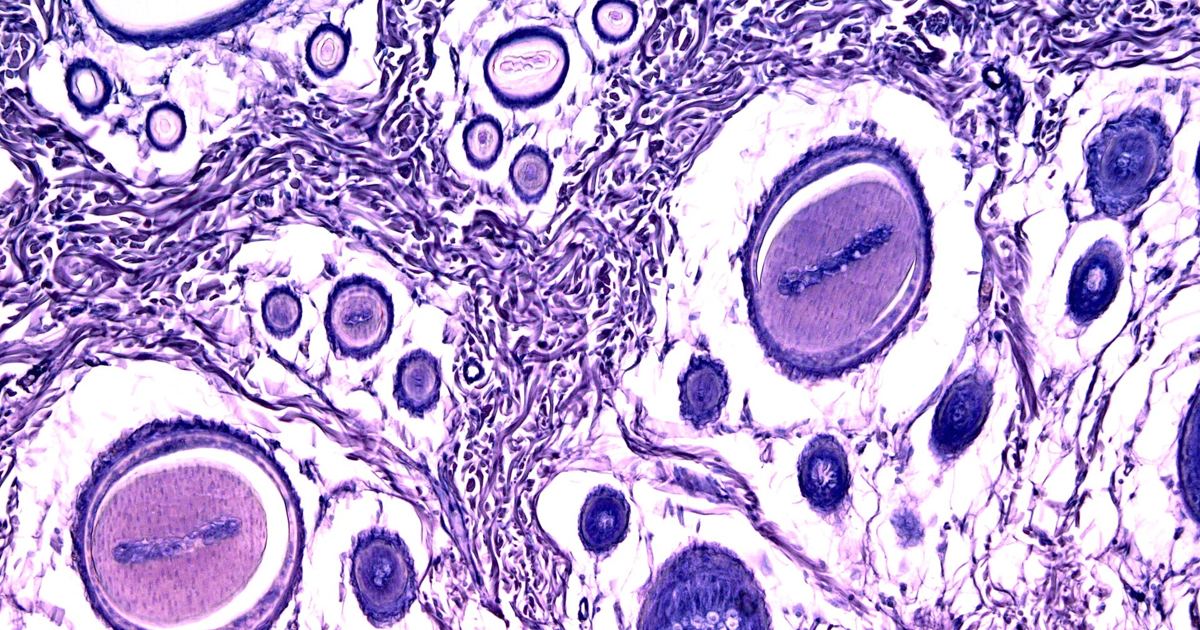
When people research melanin and skin tanning, they often assume that hair and skin produce color through identical, unified switches. However, while both processes rely on pigment-producing melanocytes and signaling molecules like alpha-melanocyte-stimulating hormone (α-MSH), hair follicles function as distinct mini-organs. Their biological regulation operates within unique microenvironments that face different controls than the surrounding epidermis. Understanding how these follicular pathways work helps explain why hair graying or depigmentation follows its own biological timeline.
Background: Skin Melanin Versus Follicular Mini-Organs
In general skin tissue, melanocytes interact with surrounding keratinocytes across broad epidermal layers, responding directly to systemic signals and external triggers. As detailed in our overview of MC1R and Skin Pigment: How Tanning Works in the Body, signaling molecules stimulate receptors on pigment cells to drive melanin synthesis. In contrast, the human scalp hair follicle operates as a self-contained anatomical structure with its own cyclic phases and localized neuroendocrine activity.
Because the follicle must continually regenerate, its pigmentary unit is tightly tied to hair growth cycles. Signals that govern cellular energy, metabolism, and protein synthesis inside the follicle can dramatically influence whether active melanocytes produce pigment. When these delicate local pathways become disrupted, follicles can lose pigment even while epidermal melanocytes remain capable of producing color, as explored in broader work on how the MC1R gene shapes skin and hair color.
Timeline of Discovery: Clues from Tuberous Sclerosis
The molecular journey connecting cellular growth pathways to hair depigmentation grew out of medical observations in rare genetic medicine. Scientists long recognized that dysregulation of mechanistic target of rapamycin complex 1 (mTORC1) plays a major role in human aging, malignancy, and specific inherited conditions.
One notable condition is tuberous sclerosis, a rare neurodevelopmental multisystemic disease defined by benign tumors, seizures, and intellectual disability. In clinical observations, patches of white hair on the scalp—a presentation known as poliosis—frequently appear as early physical signs of tuberous sclerosis. For many years, however, the direct cellular mechanisms linking mTORC1 activity to the loss of hair follicle pigment remained unresolved.
The 2023 EMBO Reports Investigation
To investigate this biological gap, researchers conducted an experimental study published in EMBO reports. Rather than relying solely on cell cultures in dishes or non-human tissue, the investigators designed their experiment using healthy, organ-cultured human scalp hair follicles. This model served as a prototypic human mini-organ system to track real-time changes in growth and melanogenesis.
The study yielded clear evidence regarding the baseline state of depigmented tissue: gray and white hair follicles exhibited elevated mTORC1 activity. This indicated that hyperactive mTORC1 signaling was not merely a bystander, but an active participant in the suppression of follicular color.
Cellular Pathways: mTORC1, TSC2, and Local α-MSH
The researchers tested the pathway by manipulating key molecular levers within the organ-cultured follicles. Their findings mapped a clear signaling chain:
- mTORC1 Inhibition: When researchers applied rapamycin to inhibit mTORC1, it stimulated both hair follicle growth and pigmentation. Notably, this repigmentation occurred even in gray or white follicles, provided they still retained surviving melanocytes.
- Intrafollicular Hormone Production: Mechanistically, the restoration of pigment driven by rapamycin operated through an increased local, intrafollicular production of α-MSH, the primary melanotropic hormone needed for melanin synthesis.
- TSC2 Regulation: To verify the inverse effect, the study performed a knockdown of intrafollicular TSC2, which functions naturally as a negative regulator of mTORC1. Reducing TSC2 unleashed mTORC1 activity, which significantly reduced hair follicle pigmentation.
Together, these steps confirmed that mTORC1 activity acts as an important negative regulator of human hair follicle growth and pigment synthesis.
Study Design, Limitations, and Patient Safety
While the findings published in EMBO reports offer fascinating insights into depigmentation biology, it is critical to interpret them within their actual scientific boundaries. This was a single laboratory study performed on organ-cultured scalp follicles outside the body. It does not represent completed human clinical trials, and laboratory manipulations of tissue culture cannot be directly translated into consumer treatments.
Furthermore, these findings highlight how tightly calibrated intrafollicular hormones like α-MSH must be. Unregulated tanning peptides sold online—such as melanotan—are unapproved, lack safety oversight, and carry unpredictable systemic health risks. They should never be used as DIY approaches for skin or hair pigment changes. Anyone noticing sudden patches of white hair, unexplained pigment loss, or skin irregularities should consult a qualified doctor or board-certified dermatologist for evidence-based clinical evaluation.
Sources
mTORC1 activity negatively regulates human hair follicle growth and pigmentation. (PubMed)
Published October 31, 2026. Sources are linked in the text. See our Medical Disclaimer.