How Acne Causes Hyperpigmentation: 2021 Study Breakdown
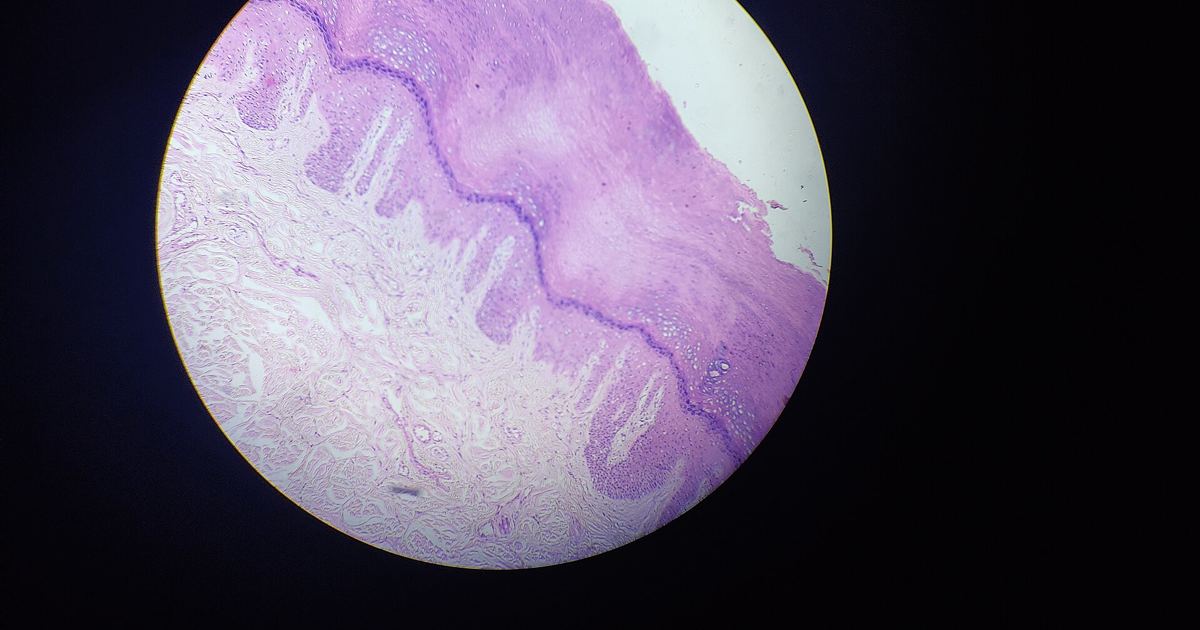
For many individuals managing acne vulgaris, the physical clearance of a pimple does not mark the end of the skin concern. Instead, resolving blemishes frequently leave behind stubborn, discolored spots. In medical terms, these lingering discolorations are classified as pigmentary sequelae. To clarify the cellular processes behind this issue, a working group of pigmentary experts published a comprehensive review examining the biological cascades that turn active acne into persistent skin discoloration.
The 2021 Expert Review: Design and Scope
The biological mechanisms connecting breakouts to dark spots were detailed in a 2021 review titled The Pathogenesis and Management of Acne-Induced Post-inflammatory Hyperpigmentation, published in the American Journal of Clinical Dermatology. This publication was organized as a literature review rather than a randomized clinical trial. To address a widespread clinical issue, the authors formed a specialized working group consisting of pigmentary experts. Their core objective was to build greater clinical awareness and provide education on how acne-related pigment alterations arise and how clinicians might approach them.
Cellular Pathways: Excess Melanogenesis and Melanin Deposition
Acne vulgaris is fundamentally classified as a common inflammatory disease. When an acne lesion develops, the surrounding cutaneous environment experiences localized inflammation. According to the review, this inflammatory response directly influences local pigment-producing cells. Specifically, the inflammatory processes of acne stimulate two critical cellular events:
- Excess melanogenesis: The biological signaling pathways prompted by acne inflammation drive melanocytes to produce surplus pigment.
- Abnormal melanin deposition: Rather than distributing pigment evenly throughout surrounding tissue, the skin retains melanin abnormally within the epidermal or dermal layers.
Together, these cellular reactions culminate in visible pigmentary sequelae, presenting either as post-inflammatory hyperpigmentation (darker brown or black patches) or post-inflammatory erythema (red or purplish vascular flushing).
Pigmentary Differences Across Skin Tones
The review emphasizes that while post-inflammatory changes can appear across all skin tones, the clinical manifestation varies markedly based on an individual's baseline complexion. In dermatologic classification, complexions are categorized using Fitzpatrick skin types.
Among patients with darker skin phototypes, designated as Fitzpatrick III-VI, the inflammatory cascade of acne most commonly triggers excess melanogenesis and abnormal melanin deposition, resulting in post-inflammatory hyperpigmentation. In contrast, post-inflammatory erythema is observed more frequently in patients with lighter skin phototypes. Even so, the authors note that both types of pigmentary sequelae can occur in any skin tone, demonstrating that inflammatory disruptions to skin homeostasis do not follow a single uniform pattern.
The Clinical Burden of Lingering Marks
A central finding highlighted by the expert panel is the substantial psychological and clinical toll imposed by acne-induced dyspigmentation. The authors noted that these pigmentary alterations can be long lasting. In routine clinical settings, these dark marks are often reported as more distressing to patients than the active acne lesions that triggered them in the first place.
Because these dark spots persist long after active papules or pustules have resolved, dyspigmentation poses both a profound daily concern for patients and an ongoing therapeutic challenge for medical providers. Effectively resolving the discoloration requires managing both the underlying inflammation and the pigmentary aftermath.
Study Limitations and Evidence Gaps
While the paper provides valuable guidance, it represents one review paper rather than a direct cellular trial. A key limitation acknowledged candidly by the authors is that much of what is currently understood about acne-related pigmentation is extrapolated from general study of nonspecific pigment deposition rather than studies focused exclusively on acne lesions. Because nonspecific pigmentary research has provided the baseline model, specific direct cellular pathways unique to acne vulgaris continue to require deeper, targeted investigation.
Protecting Skin Health and Seeking Care
Understanding that post-inflammatory marks stem directly from inflammatory cellular cascades reinforces why picking, squeezing, or applying harsh, unverified compounds to active breakouts can exacerbate tissue damage. Attempting to balance or camouflage discoloration with unregulated tanning peptides or unapproved melanocortin products purchased online poses serious systemic health risks, as unregulated products lack proven purity, clinical oversight, and safety validation.
Anyone struggling with persistent discoloration or severe breakouts should consult a board-certified dermatologist or medical doctor. A qualified healthcare provider can evaluate your skin tone, identify whether you are experiencing erythema or hyperpigmentation, and develop an evidence-backed treatment strategy based on how doctors evaluate darkened skin.
Sources
Review findings and cellular mechanisms referenced in this article are drawn from the following published medical source:
- The Pathogenesis and Management of Acne-Induced Post-inflammatory Hyperpigmentation. (American Journal of Clinical Dermatology, 2021).
Published October 31, 2026. Sources are linked in the text. See our Medical Disclaimer.