Keratinocyte Cancers vs. Melanoma: Cellular Origins Explained
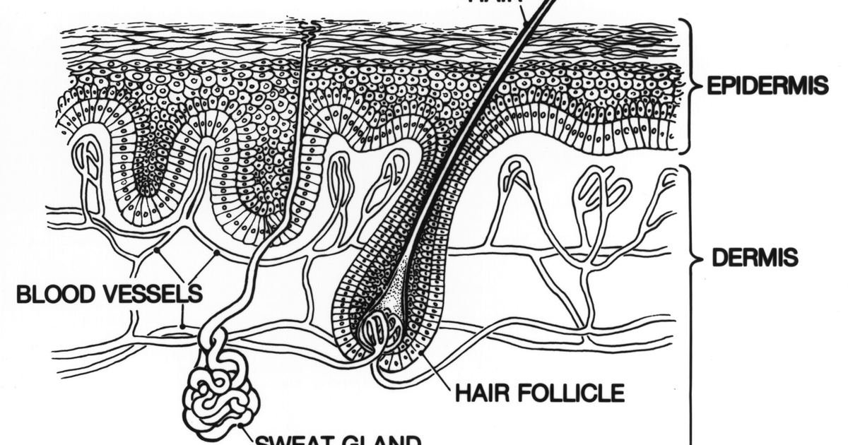
When evaluating the health consequences of sun exposure and tanning, public health authorities emphasize that skin cancer is not a single, uniform disease. According to federal public health guidance from the CDC skin cancer basics, skin cancer occurs when cells in the skin grow out of control. The exact type of malignancy that develops depends entirely on which specific cell type undergoes this uncontrolled growth. Understanding the cellular architecture of human skin provides critical insight into why different skin cancers behave, progress, and threaten health in distinct ways.
Anatomical Background: The Layers of the Skin
To understand how malignancies originate, one must first look at the basic anatomical structure of the skin. Human skin is organized into two primary layers:
- The Epidermis: The upper or outer layer of the skin. This surface layer serves as the body’s primary interface with the external environment and is the site where skin cancer begins.
- The Dermis: The lower or inner layer of the skin, positioned directly beneath the epidermis, providing deeper structural support.
Because the epidermis absorbs the brunt of environmental radiation, every primary form of skin cancer originates within this outer layer. Within this single outer shield, three specialized cell types reside, each giving rise to a distinct form of cancer.
The Cellular Foundation of the Epidermis
The epidermis contains three distinct kinds of cells that perform specific physiological tasks:
- Squamous cells: These are thin, flat cells that form the very top, outermost layer of the epidermis.
- Basal cells: These are round cells situated immediately underneath the squamous cells, occupying the lower portion of the epidermal layer.
- Melanocytes: These specialized cells reside in the lower part of the epidermis alongside basal cells. Melanocytes are responsible for producing melanin, the pigment that provides skin with its natural color.
When healthy skin is exposed to the sun, melanocytes respond by producing additional pigment, which causes the skin to darken. However, when these cells suffer significant genetic harm, their normal replication controls fail.
The Timeline of Ultraviolet Damage
The progression toward skin cancer unfolds along a clear timeline driven by radiation exposure. The vast majority of skin cancers are caused by too much exposure to ultraviolet (UV) rays, whether from natural sunlight or artificial indoor tanning beds. The biological impact of this radiation accumulates in progressive stages:
- Short-Term Injury: In the immediate term, excessive UV exposure damages skin cells directly, manifesting visibly as a sunburn.
- Cumulative Damage: Over extended periods, repeated cellular injury adds up within the tissue.
- Structural and Functional Breakdown: This sustained cellular injury leads to visible changes in skin texture and causes early skin aging.
- Uncontrolled Proliferation: Eventually, cumulative damage disrupts the mechanisms governing cell division, prompting cells to multiply uncontrollably into skin cancer.
Keratinocyte Carcinomas: Squamous and Basal Cell Origins
The two most common types of skin cancer arise from the primary structural cells of the epidermis. Squamous cell carcinoma originates in the thin, flat squamous cells forming the upper layer of the skin. Basal cell carcinoma begins in the round basal cells residing in the lower layer of the epidermis.
Public health agencies point out that basal cell carcinoma and squamous cell carcinoma are the two most common skin malignancies diagnosed. Because these cancers typically stay localized within the skin tissue for long periods, both can usually be cured when identified and addressed promptly. However, the medical interventions and surgical excisions required to eliminate them can leave noticeable scars.
Melanoma: The Lethal Behavior of Transformed Melanocytes
Melanoma represents an entirely different clinical entity because it begins in the pigment-producing melanocytes located in the lower epidermis. As outlined in the clinical timeline of how melanoma develops from melanocytes, malignant transformation in these cells carries far more dangerous systemic implications.
Although melanoma is the third most common type of skin cancer, public health officials emphasize that it causes significantly more deaths than other forms. The severity of melanoma stems from its biological capacity to invade deeper layers and metastasize. Unlike localized keratinocyte growths, melanoma frequently spreads to other parts of the body, including vital internal organs such as the brain and the liver.
Clinical Perspective and Skin Health Safety
Because excessive ultraviolet exposure from the sun or tanning beds causes most skin cancers, protecting skin cells from unnecessary radiation is the primary defense against both keratinocyte cancers and melanoma. Individuals researching tanning often encounter unregulated products sold online, such as melanotan I or melanotan II. These compounds are unapproved by regulators and carry serious health risks. Artificially stimulating pigment pathways does not replace medical photoprotection or eliminate the risks of cellular damage.
Whenever you observe changes in skin texture, persistent rough spots, or alterations in pigmented areas, consult a doctor or dermatologist. A qualified medical professional can perform a proper clinical examination, differentiate between benign changes and malignant transformations, and guide appropriate diagnostic care.
Sources
Published October 28, 2026. Sources are linked in the text. See our Medical Disclaimer.