Melanocyte Cellular Changes: How Melanoma Begins and Grows
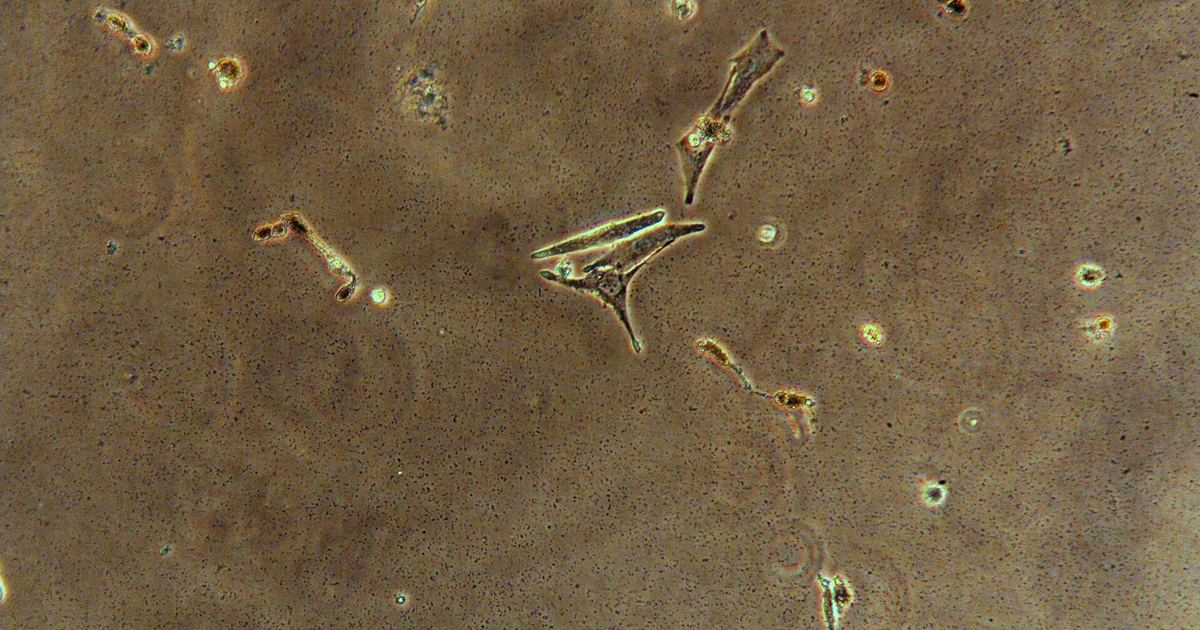
Melanoma is an aggressive form of skin cancer that originates directly inside melanocytes, the specialized cells responsible for producing skin pigment. Understanding the specific cellular transformations that enable these pigment cells to escape normal biological controls is essential for recognizing risk and understanding skin health. Rather than behaving like normal epidermal cells, mutated melanocytes develop distinct survival traits that allow them to divide continuously and spread into deeper tissues.
What Makes Melanocytes Uniquely Vulnerable to Cancerous Changes?
Under ordinary circumstances, human skin cells respond to severe ultraviolet (UV) radiation by triggering biological safety mechanisms. When cellular genetic code is broken, typical cells either stop dividing permanently (senescence) or self-destruct through programmed cell death (apoptosis). However, as explained in Melanoma: MedlinePlus Genetics, melanocytes demonstrate a much higher natural resistance to the destructive impacts of UV radiation compared to other skin cells. Even when their DNA sustains substantial damage, melanocytes are unlikely to undergo apoptosis.
Because these damaged melanocytes do not self-destruct, they persist within the skin. As these abnormal cells survive and continue to divide, they accumulate sequential mutations in the genetic pathways that govern proliferation, senescence, and cell death. Over time, this resistance to apoptosis allows rogue cells to multiply without limit or normal biological boundaries, driving the transition toward malignant melanoma.
How Does UV Radiation Trigger the Transformation from Normal Cells to Malignancy?
The primary environmental driver behind this cellular transformation is exposure to ultraviolet radiation, whether from natural sunlight or indoor tanning beds. The danger is not uniform across all types of sun exposure. According to clinical genetics data, the risk of developing melanoma is greatest after brief but intense bursts of UV exposure, rather than moderate, long-term exposure. This sudden cellular stress inflicts acute damage on cellular DNA, overwhelming repair systems.
Most cases of melanoma are sporadic, meaning the underlying genetic alterations develop over a person's lifetime rather than being passed down through families. These acquired somatic mutations remain isolated within the specific melanocytes that form the tumor. In many individuals, slight variations across multiple genes combine with environmental UV exposure to elevate risk, often presenting clinically alongside traits like a fair complexion, freckling, and having more than 25 moles on the body.
Which Specific Genes Drive Melanoma Growth and Proliferation?
As melanocytes transform, distinct genetic abnormalities disable the brakes on cellular division. Somatic mutations in the BRAF and CDKN2A genes represent two of the most frequent alterations identified in sporadic melanoma. A single significant mutation in one of these critical control genes can be enough to sharply raise the likelihood of malignant growth.
Inherited factors also alter how melanocytes respond to cellular damage:
- Familial Melanoma: Approximately 10 percent of melanoma cases occur across multiple members of the same family, stemming from inherited germline mutations present in virtually all cells.
- Key Inherited Genes: The primary genes linked to familial melanoma are CDKN2A, which helps regulate cell senescence, and MC1R, which influences skin pigmentation.
- Inheritance Patterns: These familial genetic risks generally follow an autosomal dominant pattern, meaning a single altered copy of the gene in each cell is enough to raise disease susceptibility.
- Syndromic Risk: Genetic conditions affecting the skin, such as xeroderma pigmentosum, also carry a substantially increased risk of melanocyte malignancy.
How Does Melanoma Transition from the Epidermis to Deep Tissue Invasion?
In its initial phase, melanoma usually develops within the epidermis, the thin outermost layer of the skin. Tumors can arise from an existing mole or normal skin growth that turns malignant, although many melanomas emerge as entirely new lesions. Men most frequently develop these primary tumors on the back, whereas women most commonly develop them on the legs.
When transformed melanocytes remain confined to the epidermis, the disease is localized. However, as uncontrolled cell proliferation continues, the tumor gains thickness. If the growth penetrates downward to involve multiple layers of skin, the malignant cells gain access to deeper structures and can metastasize to distant parts of the body. While the vast majority of cases involve the skin, about 5 percent of melanomas arise from melanocytes residing in other tissues, such as the eyes (uveal melanoma) or the moist mucous membranes lining internal cavities like the mouth (mucosal melanoma).
What Warning Signs Indicate That a Melanocyte Has Turned Malignant?
Because melanomas stem from pigment-producing cells, they display noticeable color and structural abnormalities as they expand. A developing melanoma frequently exhibits ragged edges and an asymmetrical, irregular outline. These lesions can vary in diameter from a few millimeters across to several centimeters. Unlike uniform benign spots, malignant melanocytic growths often present a mixture of colors, including brown, black, red, pink, blue, or white.
In the United States, melanoma is the fifth most common cancer in men (affecting 30 in 100,000 men annually) and the sixth most common cancer in women (affecting 18 in 100,000 women annually), with roughly 1 in 43 individuals developing the disease during their lifetime. Due to variations in pigmentary protection, light-skinned individuals face a 20 times greater risk than dark-skinned individuals. Furthermore, individuals who have previously had melanoma are nearly nine times more likely than the general population to develop another one. Catching these cellular shifts early is vital; approximately 90 percent of people diagnosed with melanoma survive at least 5 years. Any changing, irregularly colored, or asymmetrical mark should always be evaluated promptly by a dermatologist or medical doctor rather than self-managed or altered with unapproved cosmetic products.
Sources
Scientific details and genetic data sourced from Melanoma: MedlinePlus Genetics.
Published October 30, 2026. Sources are linked in the text. See our Medical Disclaimer.