Single-Cell Sequencing of Blocked MC1R Melanocytes: FAQ
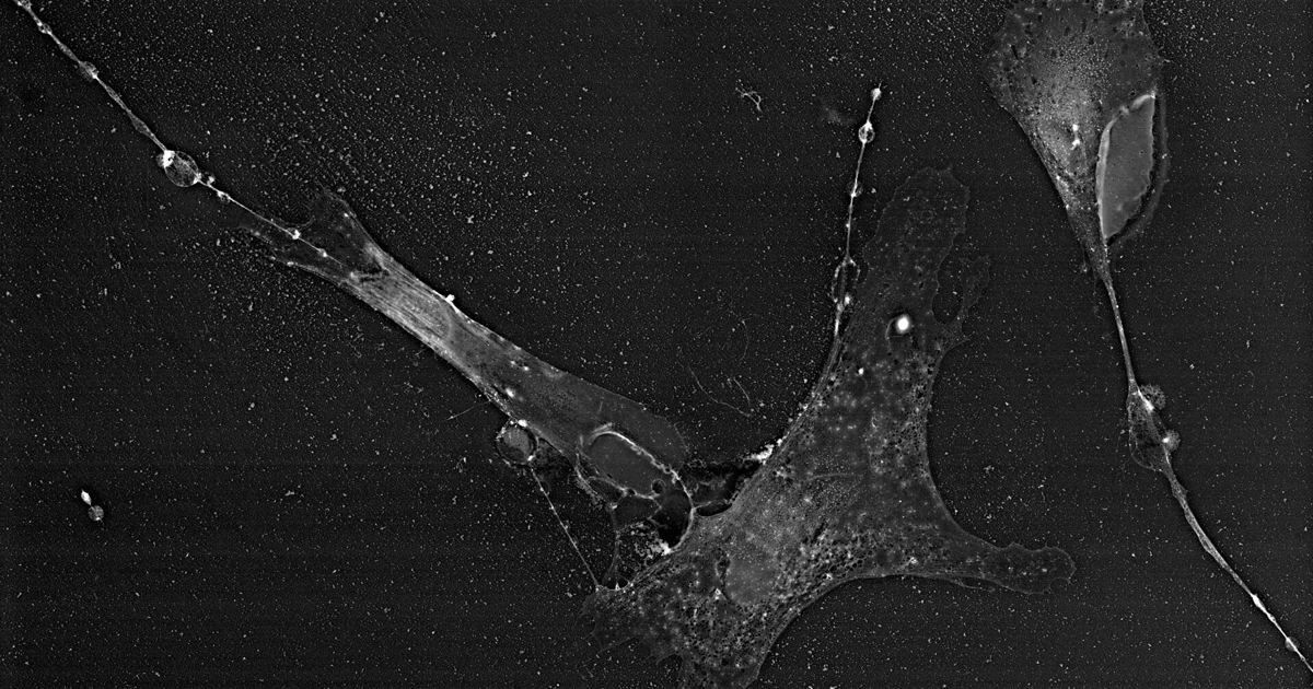
Understanding how pigment-producing cells respond when their primary surface receptors are altered is a central focus of dermatological science. A 2024 study published in Pigment Cell & Melanoma Research examined the cellular consequences of reduced receptor activity at the single-cell level. This guide reviews what the laboratory investigation discovered, how these cellular pathways interact with cellular stress responses, and why consumers researching melanocortin receptors must keep experimental data in proper clinical perspective.
What Did the 2024 Single-Cell Study Set Out to Investigate?
In humans, the red hair color trait involves an increase in red-yellow pheomelanin and a reduction in black-brown eumelanin within the skin and hair. This pigment shift is caused by diminished melanocortin 1 receptor function. While researchers have long established that receptor variants cause this phenotype and serve as a well-defined risk factor for melanoma, the exact biological mechanisms connecting reduced receptor signaling, altered pigment generation, and elevated cancer susceptibility have remained unclear.
To investigate these mechanisms, researchers conducted an analysis titled Single-cell profiling of MC1R-inhibited melanocytes. The team used single-cell RNA sequencing (scRNA-seq) on pigment cells isolated from animal models displaying the red hair color phenotype to track transcriptomic shifts on a cell-by-cell basis.
What Is the MC1R-Inhibited Gene Signature?
Single-cell sequencing allows scientists to evaluate individual cells rather than averaging gene expression across an entire tissue sample. Using this method, the study authors mapped out a distinct molecular pattern they designated as the MC1R-inhibited Gene Signature, or MiGS.
- Unidentified regulatory genes: The MiGS profile revealed a large set of previously unidentified genes that respond when receptor signaling is reduced.
- Melanogenesis shifts: The identified signature includes genes suspected of direct involvement in melanogenesis, the cellular process that governs how cells manufacture pigment.
- Malignant transformation pathways: The expression changes also implicated candidate genes that may be involved in oncogenic transformation, the process where normal cells transition toward malignancy.
By defining this signature, the researchers aimed to provide candidate targets to better understand why cells with reduced signaling behave differently when under physiological stress, as explored in broader research on MC1R and Skin Pigment: How Tanning Works in the Body.
How Does TBX3 Influence Stress Responses and Senescence?
A central finding from the Single-cell profiling of MC1R-inhibited melanocytes. investigation involved a specific candidate gene within the signature called TBX3. Cellular senescence is a natural protective arrest mechanism where damaged or stressed cells permanently stop dividing. When cells bypass senescence, they can continue replicating even after sustaining genetic or environmental insults.
The study highlighted several specific characteristics regarding TBX3 in receptor-inhibited pigment cells:
- TBX3 is recognized as an anti-senescence transcription factor implicated in melanoma progression.
- The researchers demonstrated that the TBX3 protein binds both E-box and T-box genetic elements.
- Through these binding sites, TBX3 regulates downstream genes directly associated with both melanogenesis and senescence bypass.
This provides a molecular link showing how shutting down normal receptor activity might allow melanocytes to evade normal growth-arrest checkpoints while altering the balance of pigments they synthesize.
What Are the Design Limits of This Research?
While the study reveals intricate molecular interactions, it is essential to interpret the findings within the framework of its experimental design:
- Single laboratory study: This paper represents a single study, meaning its findings must be confirmed and expanded by additional peer-reviewed investigations.
- Mouse model data: The sequencing was conducted on melanocytes isolated from mouse models carrying red hair color traits, not on living human subjects. Mouse tissue biology does not always match human cellular behavior in identical ways.
- Candidate identification: The identified genes in the MiGS profile are candidate targets for future study; the paper does not claim to provide a definitive clinical treatment or immediate diagnostic test.
Readers should avoid overstating these conclusions as direct evidence of how living human skin reacts in daily life or during synthetic chemical exposures.
How Does This Relate to Tanning Peptides and Skin Safety?
Interest in melanocortin receptors often leads people to read about experimental tanning peptides like melanotan I or melanotan II. However, fundamental laboratory research into receptor inhibition is not an endorsement of manipulating these complex pathways with illicit chemicals. Products marketed online as tanning peptides are completely unapproved by regulatory agencies and are sold without pharmaceutical manufacturing oversight.
Because melanocortin receptors regulate delicate balances involving pigment production, cell replication, and senescence bypass, interfering with these pathways carries unpredictable medical risks. Anyone noticing changing lesions, unusual pigmentation, or considering peptide use should review clinical evidence on Melanotan, Moles and Melanoma: What Case Reports Show and consult a board-certified dermatologist or qualified medical doctor for personalized advice.
Sources
Single-cell profiling of MC1R-inhibited melanocytes. — PubMed / Pigment Cell & Melanoma Research (2024)
Published October 28, 2026. Sources are linked in the text. See our Medical Disclaimer.