Squamous Cell Cancer and Sun Damage: Clinical FAQ
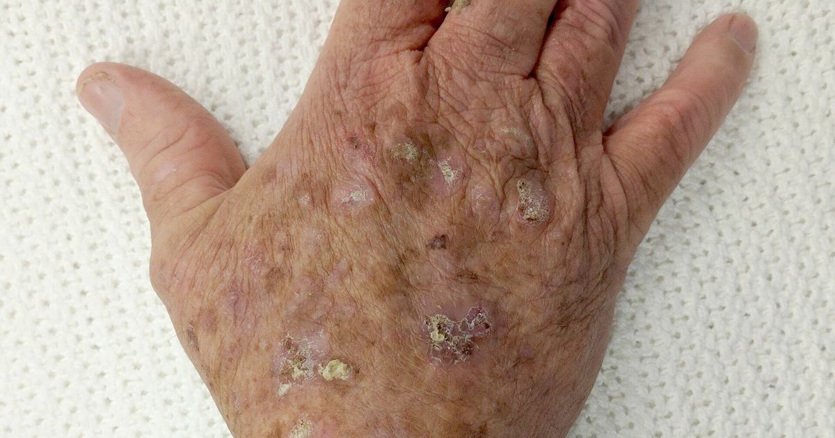
Squamous cell skin cancer represents the second most common form of cancer diagnosed in the United States. While frequently linked to ongoing UV exposure, changes in the upper skin layers often present as subtle rough spots before progressing into invasive lesions. Understanding how long-term solar radiation alters skin tissue—and why persistent, scaly growths demand prompt professional evaluation—is essential for protecting long-term dermatologic health.
How Does Chronic Sun Exposure Prompt Abnormal Squamous Cell Growth?
Squamous cell skin cancer develops in the epidermis, which is the outermost layer of the skin. Although this malignancy can arise in uninjured skin or in areas that have experienced prior injury or inflammation, the vast majority of cases develop on tissue regularly subjected to sunlight or artificial ultraviolet radiation. As detailed in the MedlinePlus Medical Encyclopedia entry on squamous cell skin cancer, prolonged exposure gradually disrupts normal cellular behavior within the epidermis.
Before an invasive cancer forms, skin damage may present as an actinic keratosis, which is recognized as a precancerous lesion capable of transforming into full squamous cell cancer. In other instances, cellular changes produce an early localized stage termed Bowen disease (squamous cell carcinoma in situ). Because Bowen disease remains confined strictly to the epidermis, the abnormal cells have not yet invaded deeper surrounding tissues. Another variation is a keratoacanthoma, a milder form of squamous cell growth that typically expands at a rapid pace.
What Signs and Symptoms Warrant Prompt Diagnostic Evaluation?
Squamous cell carcinoma most commonly emerges on sun-exposed zones, including the face, ears, neck, arms, and hands, although it may develop anywhere on the body. A primary physical sign is an expanding bump that displays a rough, crusted, or scaly exterior alongside flat reddish patches. Other prominent presentations include:
- A persistent sore that fails to heal.
- Visible alterations in an existing wart, mole, or long-standing skin lesion.
- Flat, crusted, and reddish patches that may expand larger than 1 inch (2.5 centimeters) in diameter, indicative of Bowen disease.
- Any skin spot that begins bleeding, itching, swelling, or producing localized pain.
Because untreated malignant cells carry a risk of recurring locally or spreading to lymph nodes and other distant internal organs, identifying changes in size, color, texture, or shape warrants an immediate appointment with a healthcare professional.
Who Is at the Greatest Risk for Developing Squamous Cell Lesions?
Anyone exposed to ultraviolet light can develop skin malignancies, but specific demographic and environmental factors significantly increase personal vulnerability. High-risk groups include:
- Individuals with light skin tones, red or blond hair, and blue or green eyes.
- People with a history of long-term, daily sun exposure, such as outdoor workers.
- Those who suffered numerous severe sunburns during childhood or early life.
- Older adults, as risk compounds with accumulated years of light exposure.
- Patients with compromised immune systems, particularly individuals who have undergone an organ transplant.
- Individuals with past chemical exposures, such as contact with arsenic, or a history of receiving multiple medical x-rays.
How Do Physicians Formally Diagnose and Stage Suspicious Growths?
Diagnosing skin lesions begins with a thorough physical examination by a healthcare provider, who reviews the size, shape, surface texture, and color of the abnormal tissue. Visual assessment alone is insufficient for confirmation. According to MedlinePlus guidelines on squamous cell carcinoma, a definitive diagnosis requires a skin biopsy, wherein a tissue sample is surgically removed and evaluated under a microscope by a pathologist.
Once confirmed, medical teams select a treatment plan based on the lesion's depth, anatomical location, potential spread, and the patient's general health. Most squamous cell cancers are successfully cured when diagnosed and addressed early. Standard therapeutic strategies include:
- Excision: Surgically removing the tumor tissue and closing the site with stitches.
- Mohs surgery: Systematically excising microscopic layers of skin and examining each immediately until no cancer cells remain, a method frequently utilized on high-cosmetic areas such as the nose, ears, and face.
- Curettage and electrodessication: Scraping out cancerous tissue and delivering electric current to eradicate remaining cells, reserved for shallower or smaller growths.
- Cryosurgery: Freezing superficial tumor cells to destroy them.
- Prescription topical medicines: Applying specialized skin creams containing 5-fluorouracil or imiquimod for superficial disease.
- Photodynamic therapy: Utilizing focused light activation to eliminate superficial malignant cells.
- Radiation therapy: Administered when the cancer cannot undergo surgical removal or when cells have advanced into regional lymph nodes or distant organs.
What Sun Safety Habits Offer the Best Protection?
Reducing direct exposure to solar radiation remains the most reliable strategy to prevent abnormal epidermal mutations. Dermatologists emphasize establishing consistent daily photoprotective routines:
- Apply generous amounts of broad-spectrum sunscreen that blocks both UVA and UVB rays, featuring a sun protection factor (SPF) of at least 30, to all exposed areas including the ears and tops of the feet.
- Apply sunscreen at least 30 minutes before outdoor activity, continuing use during cloudy days and throughout winter months, and reapply frequently according to package instructions after sweating or swimming.
- Limit outdoor exposure between 10 a.m. and 4 p.m., when ultraviolet radiation reaches peak intensity.
- Wear protective apparel such as wide-brim hats, long-sleeved shirts, long skirts, or full-length pants, or invest in sun-protective garments.
- Exercise caution near reflective surfaces—including sand, water, concrete, and white-painted structures—and recognize that higher altitudes cause skin to burn more rapidly.
- Completely avoid tanning salons, sun lamps, and indoor tanning booths. Spending just 15 to 20 minutes in a tanning bed presents the same level of danger as an entire day spent out in natural sunlight.
Understanding the boundaries between benign skin changes and malignant transformations is critical. Readers interested in broader diagnostic categories can explore our guide on types of skin cancer to learn how squamous cell lesions differ from basal cell carcinoma and melanoma.
Sources
Published October 28, 2026. Sources are linked in the text. See our Medical Disclaimer.