Stem Cell Migration in Skin Pigment Repair: 2021 Study Breakdown
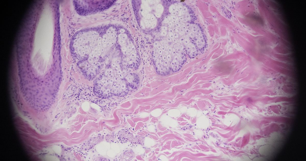
Understanding how human skin restores color after physical and environmental stress requires examining the microscopic cellular reservoirs hidden beneath the surface. When ultraviolet radiation impacts tissue, complex neuroendocrine and cellular signals coordinate pigment recovery. A scientific paper titled Skin pigmentation and its control: From ultraviolet radiation to stem cells provides critical insight into how stem cell populations travel and specialize to regenerate lost color.
The 2021 Review Breakdown: Research Scope and Limits
To evaluate these biological findings accurately, readers should consider the exact framework of the evidence. Published in Experimental dermatology in 2021, this paper (indexed under PubMed identification 33320376) is a scientific review supported by extramural grants from the National Institutes of Health and non-U.S. government funding. Rather than functioning as a direct clinical intervention trial, this paper gathers substantial laboratory discoveries to summarize epithelial and follicular pigmentation pathophysiology.
The review outlines current knowledge regarding cellular control while mapping out future intervention targets. Because it synthesizes broad laboratory literature, it does not evaluate commercial products or prove that individuals can safely manipulate these pathways on their own. Keeping these study parameters in mind prevents overstating the clinical reach of early molecular models.
The 1 Primary Receptor: MC1R and Delayed Tanning
Skin color depends directly on melanocytes, which operate as specialized pigment-producing cells. Within these cells, the melanocyte-specific microphthalmia-associated transcription factor (m-MITF) serves as the chief regulating transcription factor. Ultraviolet radiation acts as a powerful modulator of these pigment pathways, initiating what researchers call the delayed tanning response.
When ultraviolet B radiation creates DNA damage within epidermal keratinocytes, it triggers gene expression of pro-opiomelanocortin (POMC). This precursor peptide is cleaved into multiple active signals:
- Alpha-MSH: Alpha-melanocyte-stimulating hormone stimulates the melanocortin 1 receptor (MC1R) situated on melanocyte membranes, which drives m-MITF expression and accelerates melanogenesis. You can read more about this signaling cascade in our overview of MC1R and skin pigment.
- Beta-endorphin: The simultaneous release of beta-endorphin establishes a neuroendocrine circuit that promotes sun-seeking behaviors.
The review notes that the MC1R allele remains highly polymorphic, producing varied physical phenotypes across different people. Furthermore, inherited mutations along this defensive tanning pathway impair pigment production and elevate the risk of skin malignancies.
Upward Migration: Stem Cells Repigmenting the Epidermis
In conditions such as vitiligo, autoimmune destruction eliminates mature melanocytes, leaving behind stark, depigmented skin lesions. The 2021 paper explains how follicular reserves offer a biological rescue mechanism when exposed to therapeutic radiation stress. Deep within the hair bulge resides a pool of quiescent melanocyte stem cells that do not actively manufacture color under resting conditions.
When stimulated by controlled ultraviolet B phototherapy, these dormant cells awaken. Under sophisticated molecular signaling control, the stem cells undergo differentiation into active melanocytes and embark on an upward migration out of the hair bulge toward the surface. As they reach the surrounding epidermal layer, they generate perifollicular repigmentation across vitiliginous patches. Similar regenerative dynamics are evaluated in research exploring afamelanotide and UV-B for vitiligo.
Downward Migration: Hair Cycles and Stem Cell Loss
Stem cell mobilization does not move exclusively upward. Under routine physiological conditions, follicular melanocyte stem cells participate in the cyclic rhythm of the hair cycle. During these normal cycles, the cells activate, differentiate, and undertake downward migration into the hair bulb to supply fresh pigment to emerging hair fibers.
Over time, the progressive, natural loss of these stem cell reserves leads to physiological hair greying. The 2021 review highlights that acute physical stress can severely accelerate this greying process. Specifically, acute stress triggers sympathetic nerve activation that drives rapid hyperproliferation of melanocyte stem cells, prematurely emptying the hair bulge reservoir and permanently exhausting follicular pigment potential.
Clinical Guidance and Laboratory Evidence
The intricate mechanisms described in Skin pigmentation and its control: From ultraviolet radiation to stem cells show that skin repigmentation is tightly regulated by genomic pathways, cellular migration, and specialized stem cell niches. Disruptions anywhere in this chain can alter protective barriers or elevate cancer risks. Anyone noticing unexpected pigment changes, hair depigmentation, or sun sensitivity should consult a board-certified dermatologist or medical doctor rather than attempting self-directed treatments.
Consumers should also recognize that tanning peptides, melanotan variants, or related synthetic chemicals sold online are unapproved, unregulated drugs. These illicit compounds bypass essential clinical oversight, present unknown biological hazards, and carry serious health risks. Legitimate dermatological therapies rely on rigorous clinical verification, precise phototherapy protocols, and professional medical supervision.
Sources
Published October 28, 2026. Sources are linked in the text. See our Medical Disclaimer.