Surgical Suturing and Eruptive Moles: Comparing Trauma Triggers
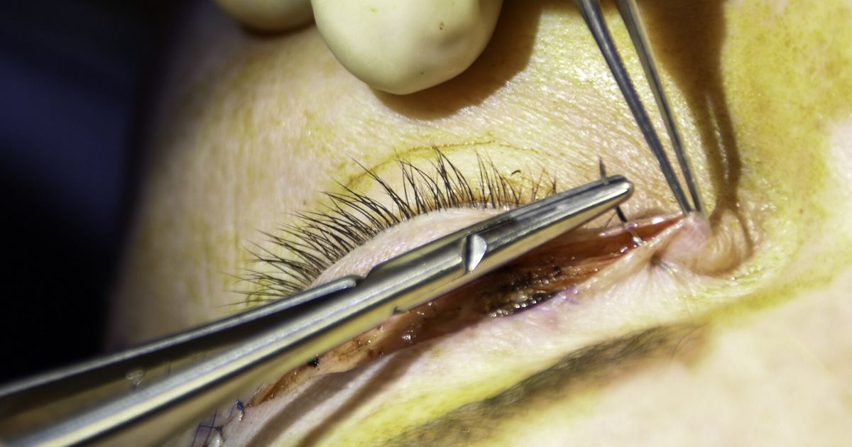
When new pigmented lesions develop on the body, patients and clinicians usually evaluate classic triggers such as ultraviolet radiation, baseline genetic traits, or internal hormonal changes. However, clinical researchers have documented unusual physical circumstances where localized mechanical injury can prompt the appearance of new moles. Understanding how skin disruption interacts with melanocytes provides valuable context for evaluating unusual skin marks and reinforcing why any new or shifting pigmentation should be examined by a dermatologist.
Standard Nevogenesis vs. Trauma-Induced Pathways
The formation of melanocytic nevi, known medically as nevogenesis, is recognized as a complex biological process. Standard medical models identify several primary drivers of nevogenesis, including genetic factors, hormonal influences, and exposure to UV-radiation. These mechanisms stimulate melanocytes to cluster, producing common acquired moles across various areas of the skin.
In contrast to these familiar systemic or environmental triggers, mechanical trauma has been identified as a triggering factor for an alternative pathway of nevogenesis. When skin tissue experiences physical disruption, the local cellular environment changes substantially during healing. This mechanical pathway demonstrates that pigment cells can respond directly to physical tissue insult, altering how doctors interpret new spots appearing near sites of prior physical damage.
Systemic Eruptive Nevi vs. Localized Suture-Induced Nevi
To understand trauma-related mole outbreaks, clinicians contrast them with traditional eruptive melanocytic nevi. Classic eruptive nevi typically emerge in broad, systemic contexts. Medical literature links these widespread eruptions either to profound immunosuppression or to extensive blistering disorders.
In those blistering or immunosuppressive conditions, the sudden appearance of multiple nevi is believed to stem from widespread disruption of the dermo-epidermal junction. As the skin repairs this boundary, quiescent pigment cells undergo rapid proliferation during the re-epithelization phase. Suture-induced nevi share this core cellular mechanic but differ in scale. Rather than scattering across broad anatomical zones under systemic immune compromise, suture-induced lesions remain strictly confined to the precise physical path of surgical intervention.
The 2016 Case Report: Study Design and Key Observations
A focused medical investigation detailed this phenomenon in the Journal of dermatological case reports under the title Surgical suturing-induced melanocytic nevi. A new type of eruptive melanocytic nevi?. Because this publication is a clinical case report rather than a large randomized trial, its scope is inherently focused and specific.
The researchers documented two patients who developed three melanocytic nevi de novo. In each instance, these new lesions formed strictly along the direction of surgical suturing following a surgical operation performed for an unrelated medical reason. Rather than developing randomly or across sun-exposed skin, the nevi tracked the physical lines created by the needle and suture material during wound closure.
Comparing Clinical Features: Dermoscopy and Histology
When evaluating unexpected spots along a surgical incision, determining whether the tissue is benign or abnormal requires formal clinical assessment. The authors of the 2016 study evaluated the three lesions using both surface examination and tissue analysis:
- Dermoscopic presentation: The spots displayed special dermoscopic characteristics distinct from ordinary flat scar tissue.
- Histological structure: Microscopic tissue examination revealed the classic architectural and cellular features of acquired melanocytic nevi.
- Linear orientation: The lesions arose de novo and followed the physical trajectory of the suturing thread used to close the surgical site.
These findings confirmed that despite their unusual origin and linear grouping along wound margins, the physical growths were true melanocytic nevi resulting from local pigment cell activity during wound repair.
Differential Diagnosis: Moles vs. Scar Pigmentation
A primary lesson from this research involves the differential diagnosis of healing incisions. New pigmentation developing within or alongside a scar can present a diagnostic dilemma. In regular post-operative recovery, dark marks may reflect ordinary post-inflammatory healing or simple pigment deposition within scar tissue.
However, because healing tissue can occasionally harbor atypical growths or unexpected melanocytic changes, clinicians emphasize that surgical suturing-induced nevi should be specifically included in the differential diagnosis of any new pigmentation emerging within a scar. A professional evaluation with dermoscopy and biopsy helps clinicians differentiate benign suture-line nevi from other pigmentary anomalies or malignant changes, a critical distinction explored further in clinical discussions of moles and melanoma.
Study Limitations and Clinical Realities
While the findings from this Journal of dermatological case reports article provide fascinating biological insight, readers must keep its design limitations in mind. The study describes only two patients and three individual nevi. Because of this small sample, the report cannot establish general incidence rates, determine whether specific suture materials carry higher risks, or predict how often surgical patients might experience this reaction.
What the study does confirm is that disruption of the dermo-epidermal junction during surgical repair can occasionally awaken quiescent pigment cells during re-epithelization. Anyone noticing new, dark, or changing spots along a surgical scar should avoid self-diagnosis and seek a full clinical exam from a board-certified dermatologist.
Sources
Surgical suturing-induced melanocytic nevi. A new type of eruptive melanocytic nevi? - PubMed
Published October 31, 2026. Sources are linked in the text. See our Medical Disclaimer.